
Abstract
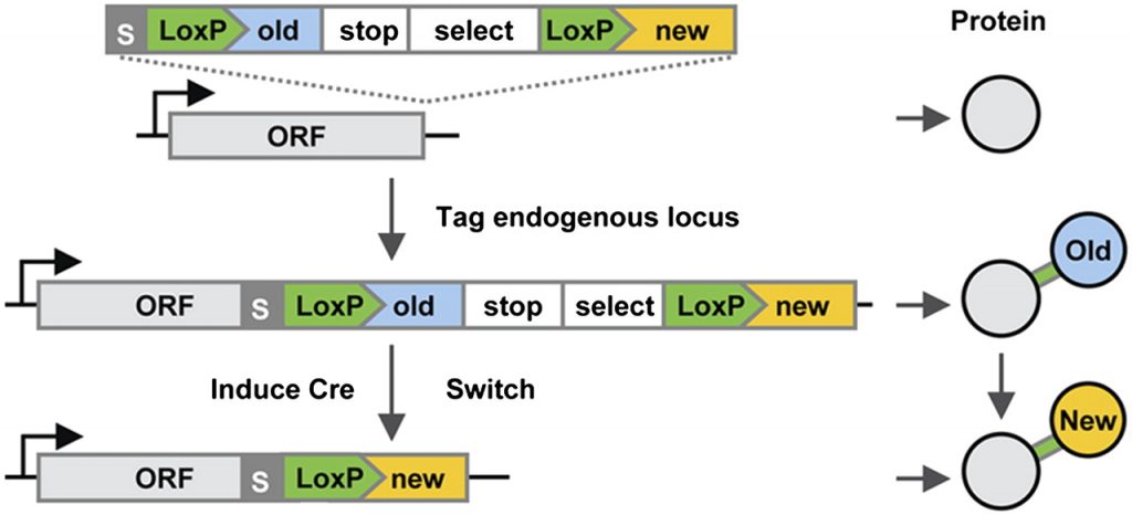
A new method of gene tagging (tag fusion tagging [MFT]) for Neurospora crassa and Magnaporthe oryzae is demonstrated. Translational fusions between the hygromycin B resistance gene and various markers are inserted into genes of interest by homologous recombination to produce chromosomally encoded fusion proteins. This method can produce tags at any position and create deletion alleles that maintain the N- and C-terminal sequences. We show the utility of MFT by producing enhanced green fluorescent protein (EGFP) tags on proteins localized to nuclei, spindle pole bodies, septal pore plugs, Woronin bodies, developing septa, and the endoplasmic reticulum.
Cell Tags and Markers Research Tools
Tags and Cell markers recombinants are a unique set of proteins located on the cell surface that allow the identification, classification and visualization of cells with antibodies; these antibodies can be directed against a single target or multiple targets depending on the type of cell and the unique set of cell markers present. Organelle markers, also known as subcellular markers, are proteins that are specific to an organelle; these allow the identification and visualization of these structures with antibodies.
Epitope tags are short peptide sequences, usually made up of 10 to 15 amino acids, designed to create a molecular identifier for a protein. An epitope tag can be placed anywhere within a protein but is usually placed at the C-terminus or the N-terminus to minimize alterations of tertiary structure that can alter protein function.
By introducing a unique sequence of amino acids into a recombinant protein, antibodies against the corresponding epitope tag can be used to track protein expression and visualize the protein in tissues, cells, and organelles.
We offer a comprehensive portfolio of antibodies, proteins, and assays, to detect and quantify cell labels and markers, that exhibit high specificity, activity, performance, and reproducibility in a variety of techniques, including Western Blot (WB), Immunohistochemistry (IHC). ), immunofluorescence (IF), immunocytochemistry (ICC), and ELISA.